Structure and functions
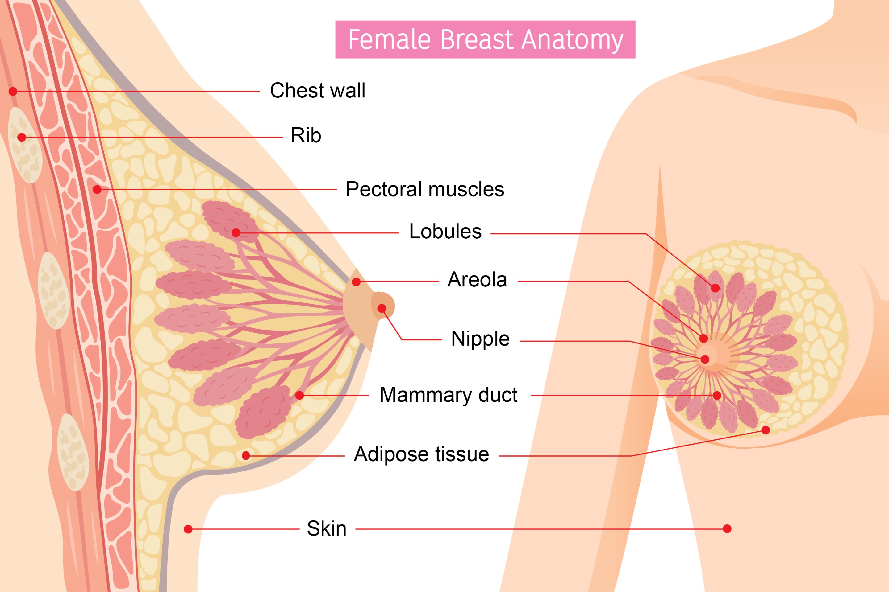
Breasts are made up of breast tissue and fat, and are held in place between skin and chest muscle by connective tissue.
Breast tissue consists of a complex network of lobules (glandular tissue that produce milk) and mammary ducts (channels that drain milk from the lobules to nipple openings during breastfeeding). Lobules and ducts are arranged in a pattern that looks like bunches of grapes.
The colour of Nipples
Nipples and areolas come in different sizes and colours, from light pink to dark brown. The colour of nipples usually relates to skin colour. It is absolutely normal to have some hairs growing around the nipples.
The skin conditions
The skin tissue of breasts is the same as the skin tissue of the rest of the body. When there is skin condition occurs (such as itchiness, flaking and thickening), patients often concern that it may represent underlying breast cancer. These conditions can be designated into malignant, infectious and inflammatory categories. Patient should seek medical advice early. Clinical examination and sometimes skin biopsy are used to evaluate these conditions.
The difference in breast tenderness between pre-menstrual cycle and others
Breast pain is a common condition. It can be cyclical or non-cyclical. Cyclical breast pain occurs a few days to a week before the menstrual cycle; it could be related to hormonal change. Non-cyclical breast pain does not occur regularly. It can be caused by injury, inflammation or previous breast surgery. Although it is unlikely to identify the exact cause of breast pain, pain is considered to be unrelated to breast cancer.
Is it common to have breast asymmetry?
It is common for breasts to be somewhat different in size. They are sisters, not twins.
Will eating chicken wings increase the risk of getting breast cancer?
There is a saying about eating chicken wings increases breast cancer risk. However, there is no scientific evidence to support this currently.
How many diagnostic imaging tests are available for breasts screening? What are they?
There are two screening breast imaging tests to identify breast cancer at early stage when it is more curable; this includes mammogram and breast ultrasound.
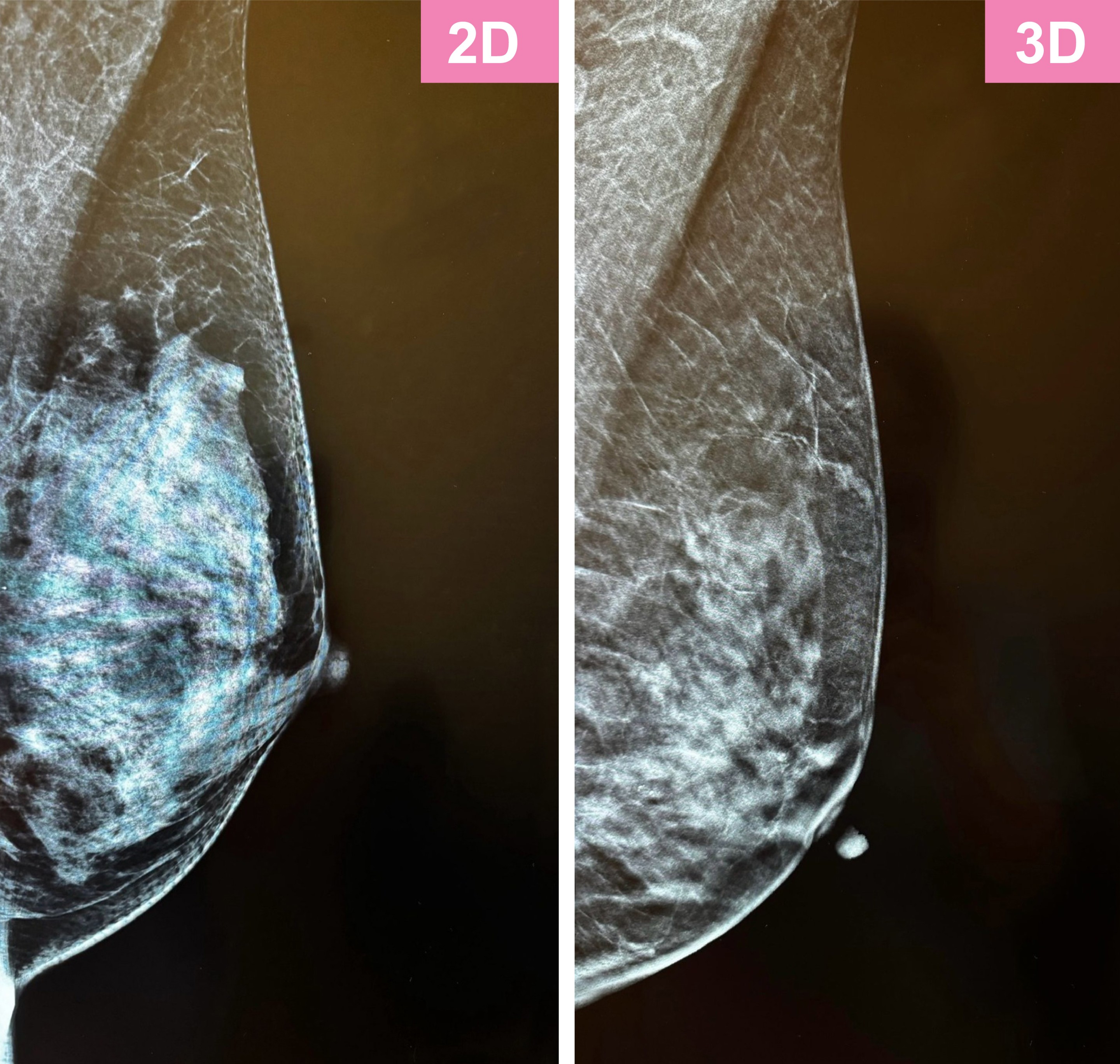
Mammogram is a low-dose X-ray exam of breast tissue. During a mammogram, your breast is placed onto the X-ray machine. It will be compressed while the X-rays are taken. Breast compression is necessary for a mammogram to hold your breast still and minimize movement, which can affect image quality. Compression also evens out the shape of your breast so that the X-rays can travel through a shorter path. This allows for a lower radiation dose and improves the image quality. Screening mammogram is recommended for women at the age of 40 and above.
Tomosynthesis, also known as 3D mammogram, uses X-ray to create a three-dimensional imaging of the breast. It has been shown to increase breast cancer detection as compared to traditional 2D mammogram. In addition, it is more comfortable as it places less pressure on breast.
Breast ultrasound uses high-frequency sound waves to produce two-dimensional image of breast tissue. In the past, ultrasound was not considered as a breast screening tool. But as dense breast is common among Hong Kong women and mammogram is less effective in breast cancer detection in dense breast, ultrasound should be considered as an additional screening option. When mammogram and breast ultrasound are used together, we can have a comprehensive assessment of breast tissue.

> If you have any questions or concerns, please feel free to schedule a consultation appointment with our surgeons.


